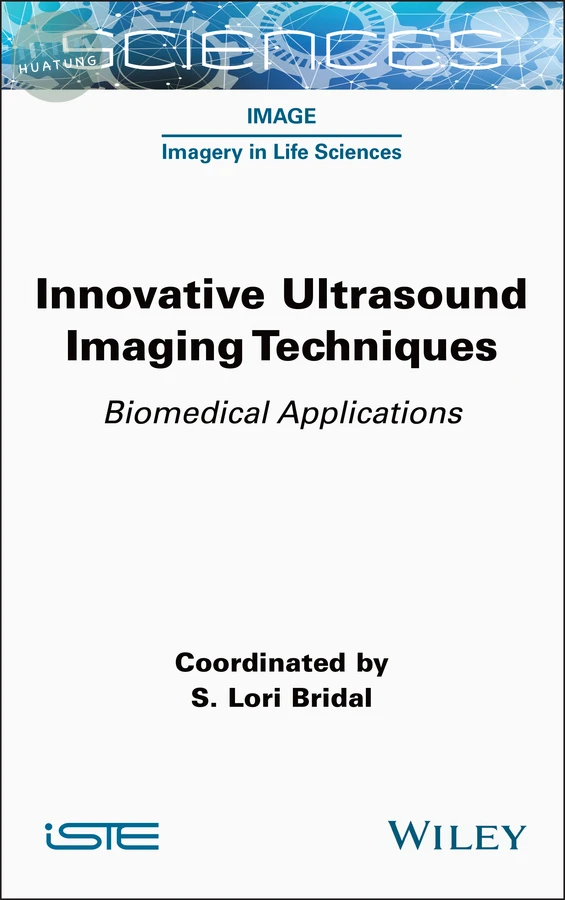
Innovative Ultrasound Imaging Techniques
$4897元 定價: $5441 9折 可訂購
簡介
This book provides an understanding of ultrasound imaging principles and how the field is evolving to better probe living systems. Today, widely-used imaging systems visualize structures and blood flow within the body in real-time. Signal analysis, hardware and contrast agent innovations are extending the capacity of ultrasound to assess tissue elasticity, to enable three-dimensional viewing of moving structures and to detect vessels smaller than the wavelength-limited resolution. Techniques are also being designed so that we are less impeded by bones in the sound path, as well as to combine light and sound to detect optically-absorbent structures within the body.
After an introductory chapter reviewing the key basic concepts, each chapter presents a detailed explanation focusing on a specific set of key principles and then shows the related techniques in each domain that are currently being refined to evaluate living systems in greater depth.
目錄
Chapter 1. Principles of Ultrasound Imaging and Signal Analysis 1
S. Lori BRIDAL
1.1. Introduction: probing living systems with ultrasound 1
1.2. Ultrasound probes 4
1.3. Longitudinal ultrasound waves 11
1.4. Pulse-echo ultrasound 17
1.5. Forming an image 25
1.6. Imaging modes to further probe living systems 30
1.7. References 34
Chapter 2. Transducers, Imaging Systems and Image Formation 37
Enrico BONI, Alessandro RAMALLI, Alessandro Stuart SAVOIA and Piero TORTOLI
2.1. Ultrasonic transducers 37
2.2. Image formation techniques 51
2.3. Advanced imaging systems 60
2.4. References 63
Chapter 3. Shear Wave Propagation and Probing Tissue Mechanical Properties 71
Stefan CATHELINE and Bruno GIAMMARINARO
3.1. Introduction: a historical perspective of palpation 71
3.2. Sound waves in human tissue: the liquid man 75
3.4. The shear wave source problem 79
3.5. Elastography beyond ultrasound 85
3.6. References 87
Chapter 4. Doppler Ultrasound and Flow Mapping 93
Alfred C.H. YU
4.1. Continuous-wave (CW) Doppler 94
4.2. Pulsed Doppler 100
4.3. Color flow imaging (CFI) 108
4.4. New flow mapping techniques 114
4.5. Summary 119
4.6. References 120
Chapter 5. Cardiac Ultrasound Imaging 123
Konstantina PAPANGELOPOULOU, Marta ORLOWSKA, Sjoerd NOOIJENS and Jan D’HOOGE
5.1. Introduction to cardiac ultrasound 123
5.2. High-frame-rate imaging (in 2D) 135
5.3. Volumetric imaging 140
5.4. References 148
Chapter 6. Ultrasound Contrast Agents: Microvascular Characterization 159
Simona TURCO, Peiran CHEN, Andrej LYSHCHIK, Ahmed EL KAFFAS and Massimo MISCHI
6.1. Introduction 159
6.2. Ultrasound contrast agents and their acoustic response 162
6.3. Analysis of UCA kinetics 167
6.4. Current challenges and future perspectives 188
6.5. References 192
Chapter 7. Resolution Limits and Super-Resolution Imaging 207
Vincent HINGOT and Olivier COUTURE
7.1. Introduction 207
7.2. Ultrasound localization microscopy 218
7.3. Topics in ultrasound localization microscopy 227
7.4. Perspectives and challenges 233
7.5. References 240
Chapter 8. Sources of Image Degradation and their Correlation in Single-sided Ultrasound Imaging of Heterogeneous Tissues 249
Guillaume RENAUD, Danai E. SOULIOTI and Gianmarco PINTON
8.1. Introduction 249
8.2. Sources of image degradation 250
8.3. Correction methods 265
8.4. Current limitations for correcting the sources of image degradation and future 277
8.5. References 278
Chapter 9. Tomography and Spectroscopy: Photoacoustics 285
Théotim LUCAS and Jérôme GATEAU
9.1. Combining ultrasound with optical waves 285
9.2. Generation and tomographic detection of ultrasound in photoacoustic imaging 288
9.3. Multispectral photoacoustic imaging 301
9.4. Conclusion and perspectives 308
9.5. References 309
Glossary 315
List of Authors 331
Index 335